Design


Overview

How we design Doara?
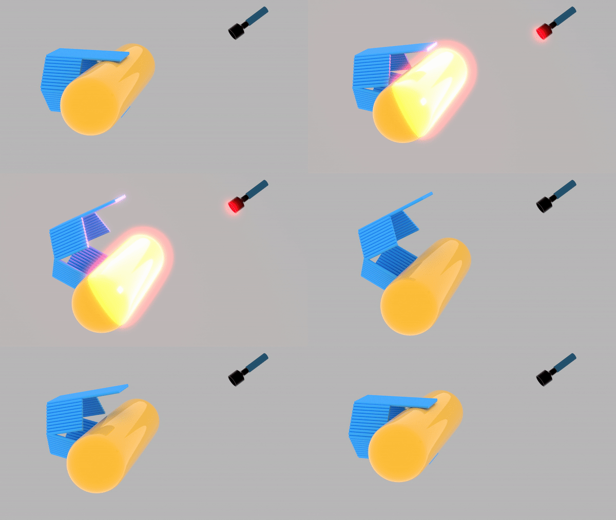
We have engineered a DNA Origami-Au nanoRod Assembly (Doara) that is capable of being remotely actuated to open or close by gold nanorod (AuNR). The AuNR is positioned at the center of the Doara, keeping it in a closed conformation. Exposure to near-infrared irradiation induces heat generation by the AuNR, which in turn denatures the heat-sensitive DNA on its surface[1], triggering the opening of the Doara.
The Doara has been designed with an internal cargo compartment and equipped with aptamers for the specific binding and carriage of molecular payloads, such as chemokines[2]. This design confers the potential for the remote-controlled delivery of therapeutic agents, thereby enabling the elicitation of targeted immune responses in vivo[3].


DNA origami

The details of our DNA nano structure
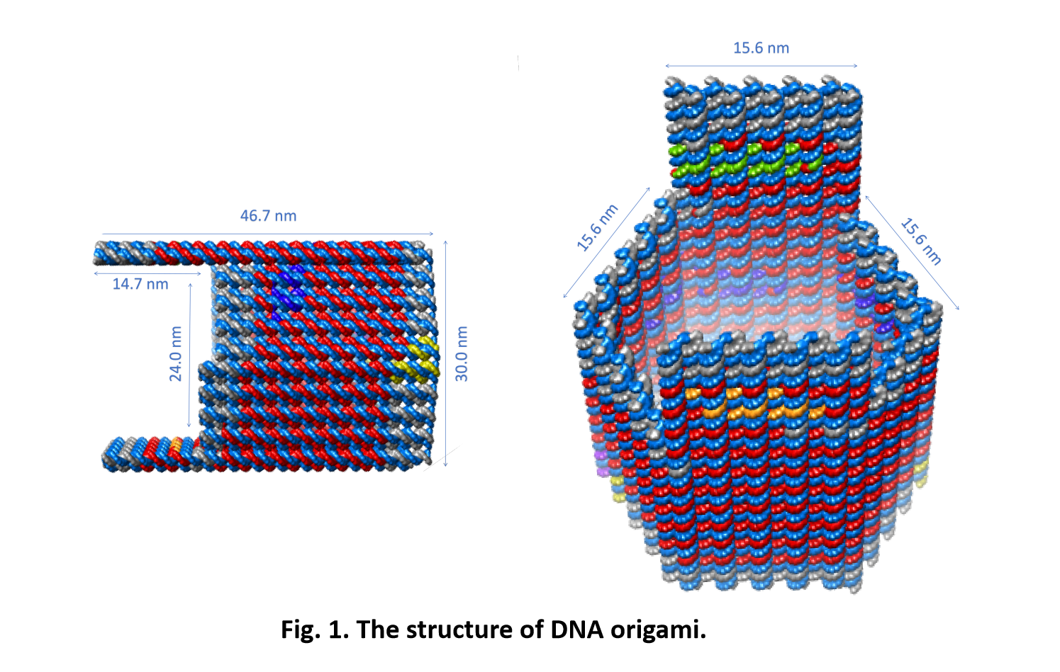
Our DNA origami is designed as a hollow hexagonal prism, with dimensions of 15.6 nm in length and 46.7 nm in height for each lateral edge, comprised of a single layer DNA.
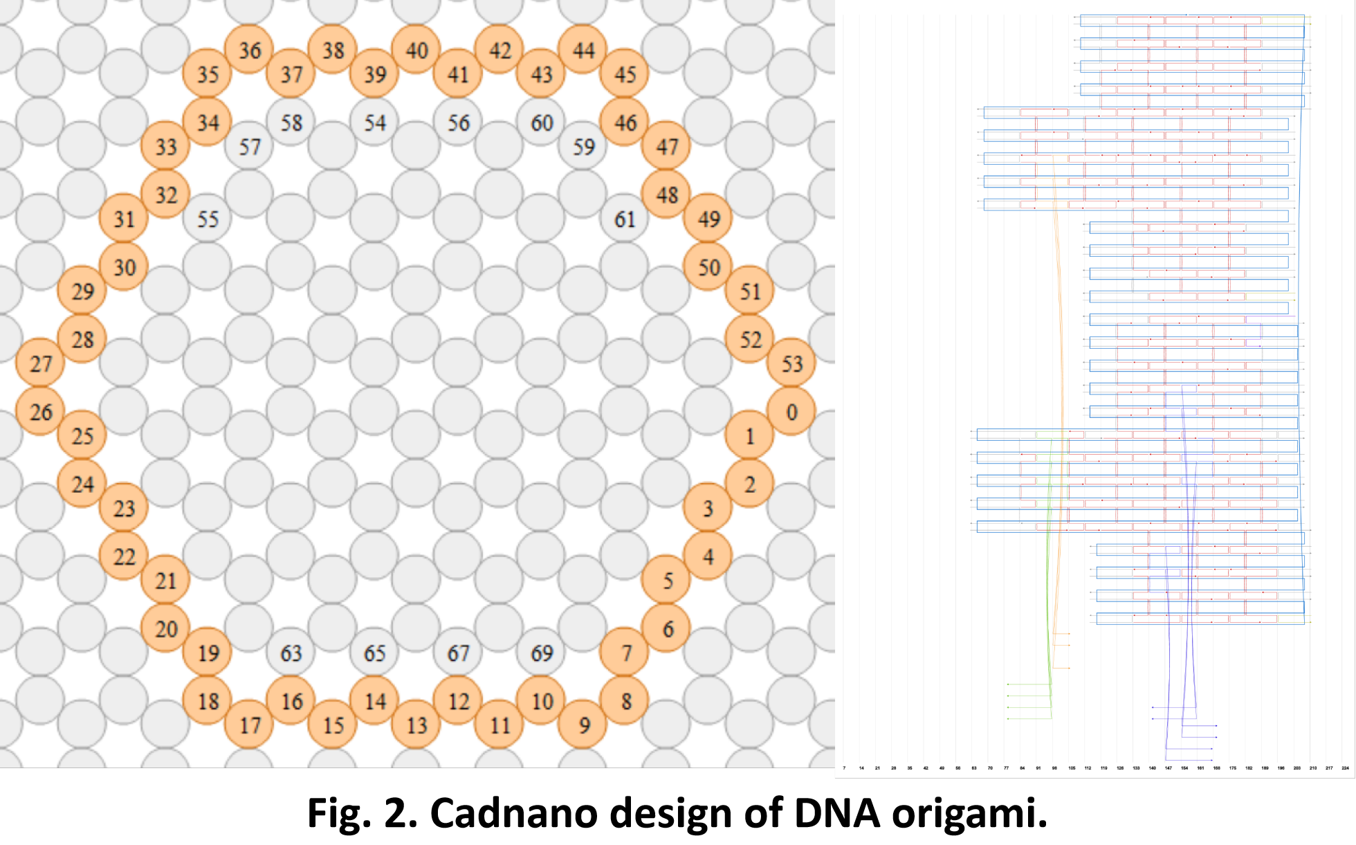
We employed the caDNAno software to design our DNA origami and it was constructed by 169 staple strands[4][5]. The scaffold for the DNA origami was 7249 nt M13mp18 plasmid, with an average staple length of 37 nt.
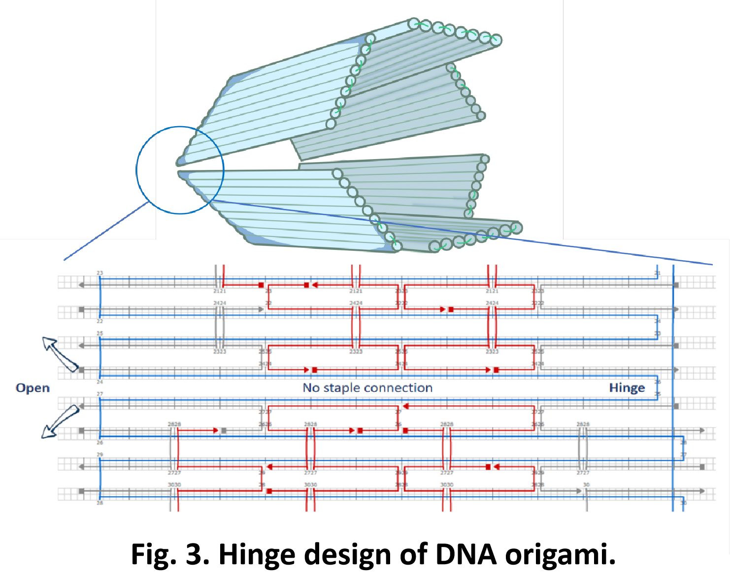
The structure features a central division and a hinge that interlinks the upper and lower portions of the prism, endowing the origami with the capability to undergo opening and closing motions[6].

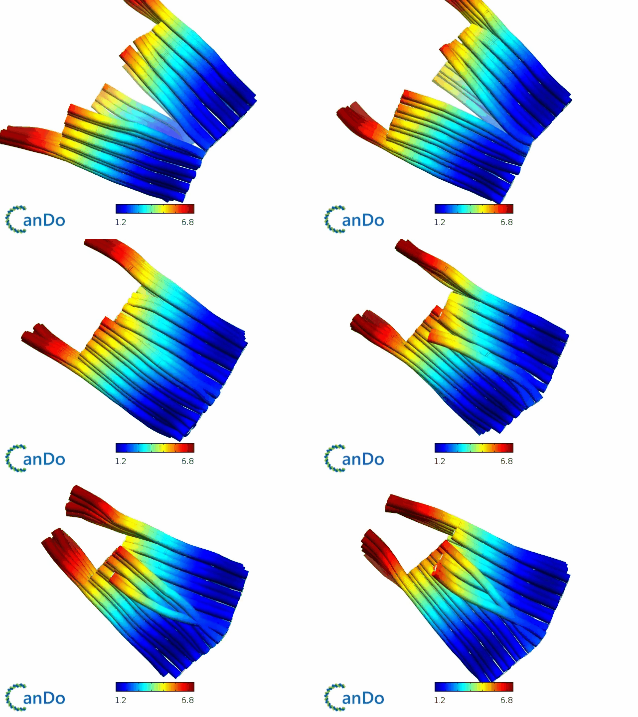
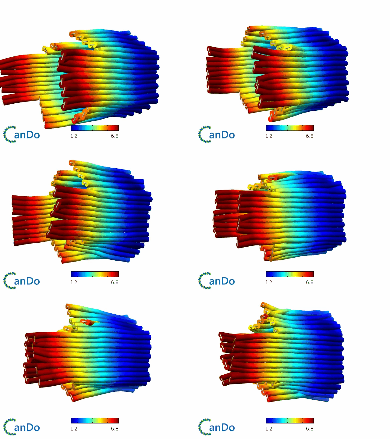
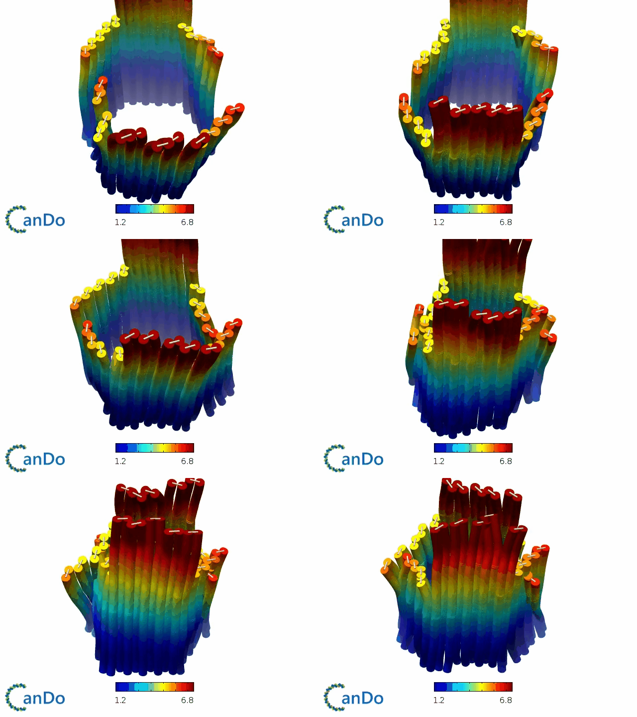
Simulation video of DNA origami drawn by Cando [7].

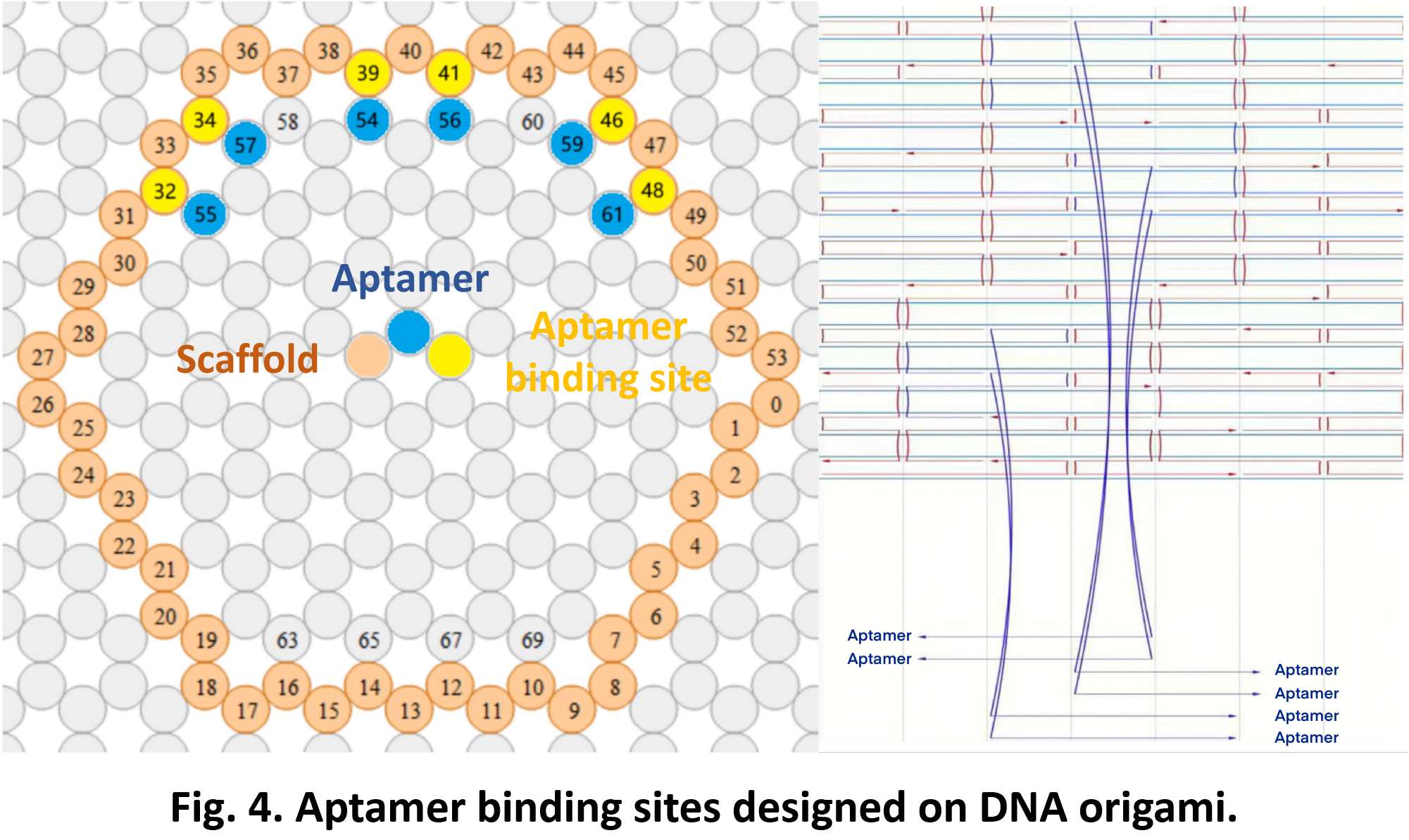
The interior of the structure has been functionalized with aptamers for the carriage of molecular payloads.[8]
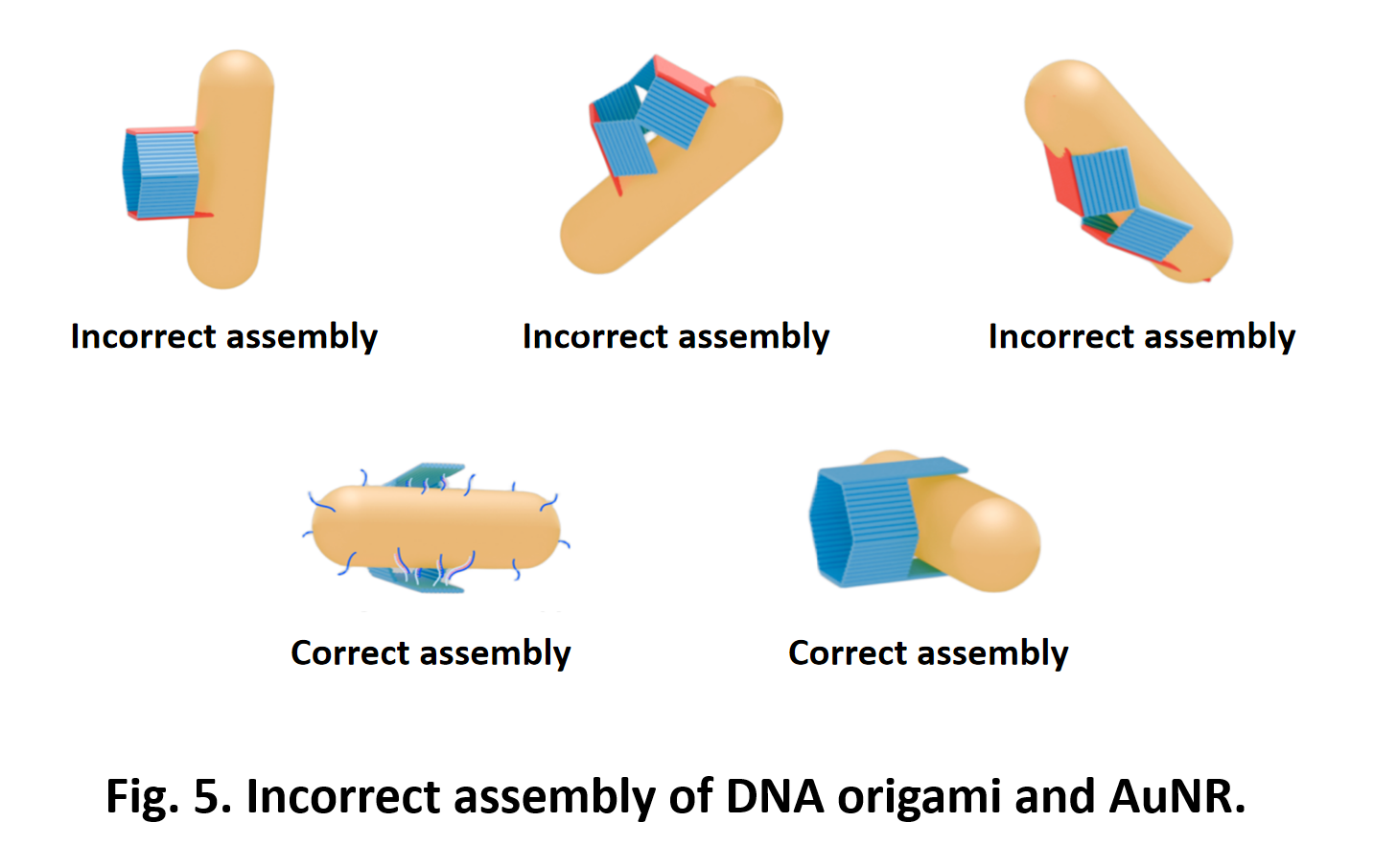
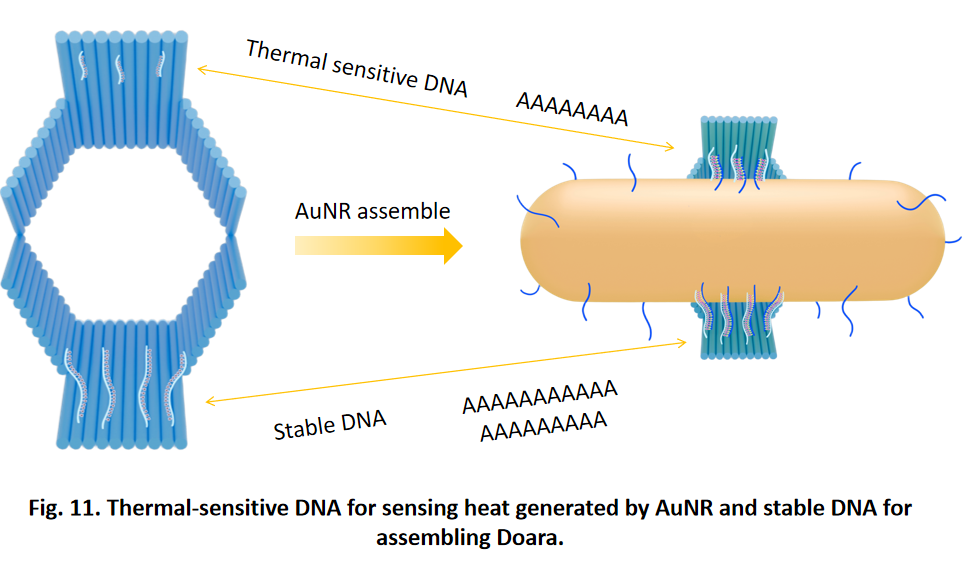
In order to ensure AuNR can be combined to DNA origami in a transverse way, We have designed extended upper and lower layer and the DNA strand which connected to AuNR is designed inside the DNA origami to avoid incorrect assembly.
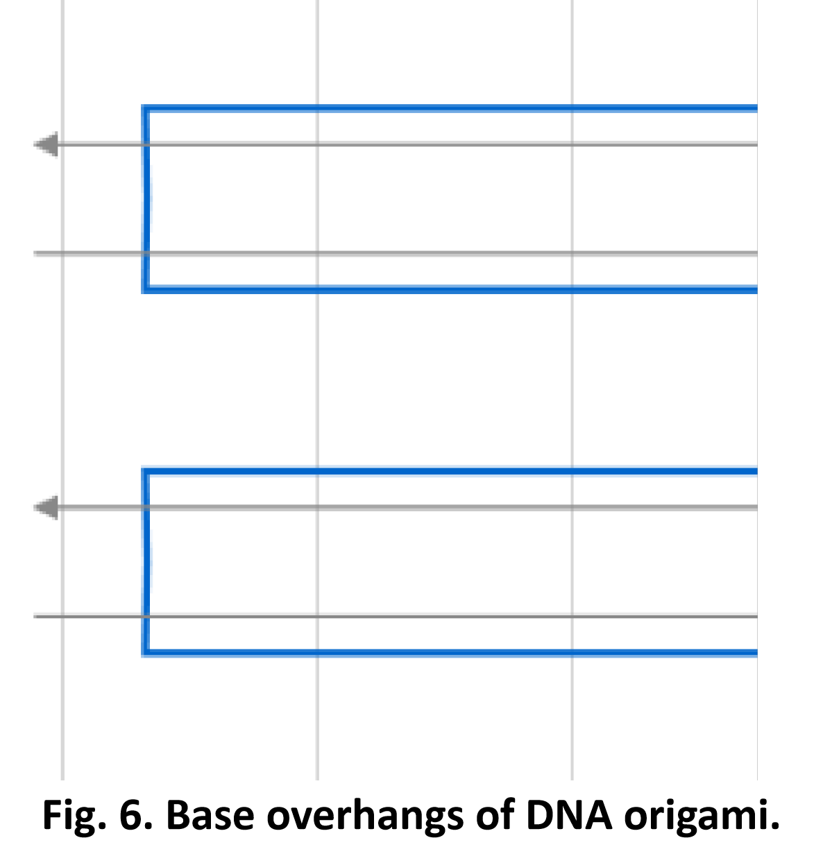
We have modified 3nt-long base overhangs on the most-lateral helices of DNA origami to prevent aggregation.


AuNR

The size of AuNR and DNA origami assembly
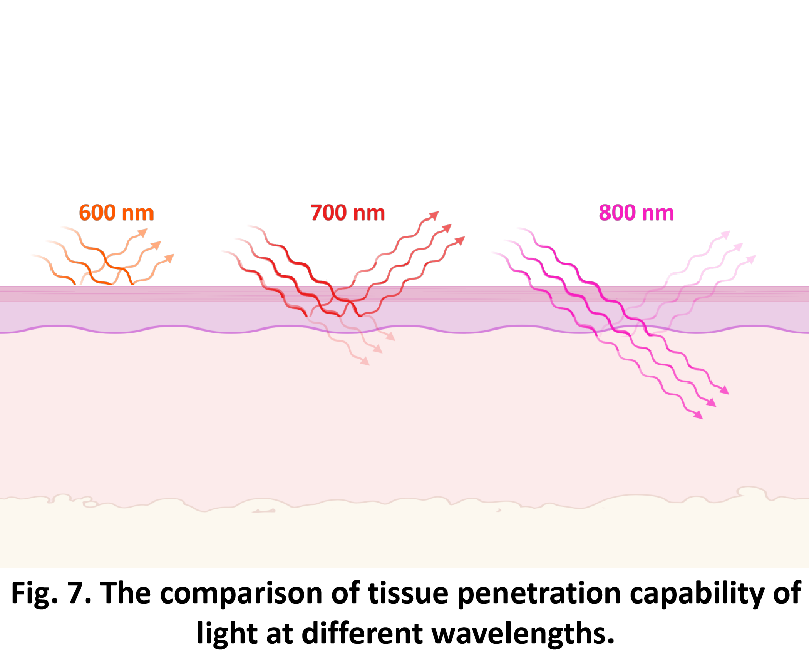
This wavelength is particularly advantageous due to its superior tissue penetration capabilities. Such a property enables the remote control of DNA origami within biological systems, potentially allowing for the manipulation of these nanostructures inside the body without causing damage to the surrounding tissues.[10]
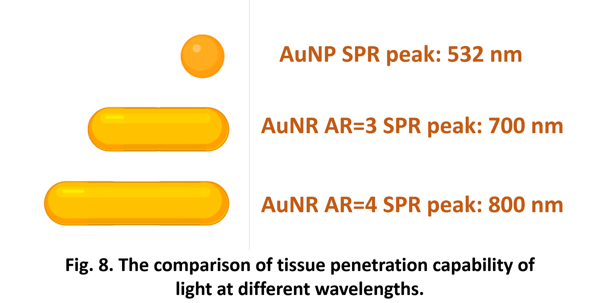
We have chosen AuNR with a aspect ratio of 4 and the optimal wavelength for its surface plasmon resonance (SPR peak) is 800 nm, which is in the near-infrared region. [11]
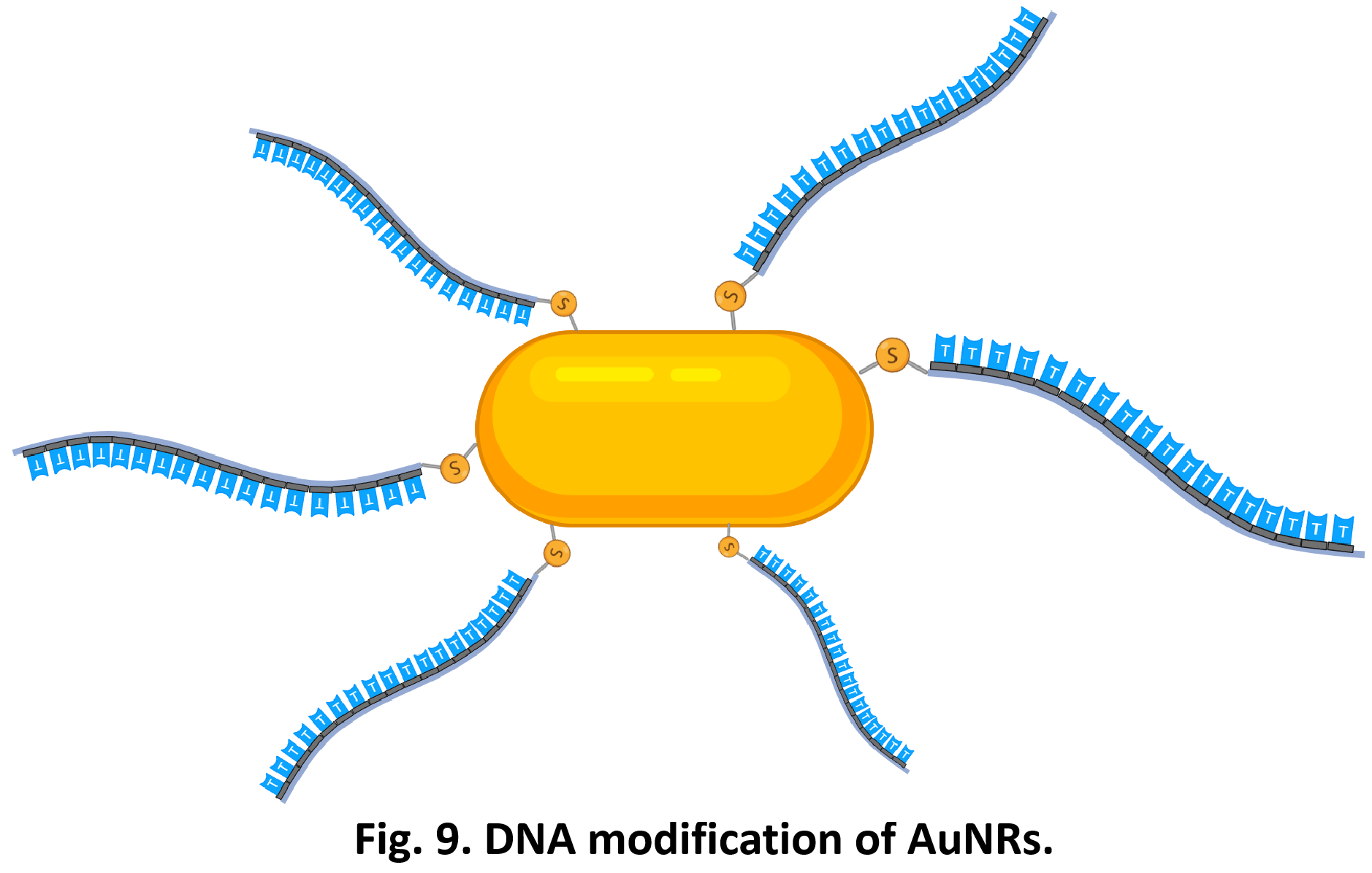
We employed thiol-modified DNA, which can coordinate with AuNRs through Au-S bonding, thereby conjugating the DNA onto it. The DNA sequence conjugated to the AuNRs is 5' HS-TTTTTTTTTTTTTTTTTTTT-3'.[12]
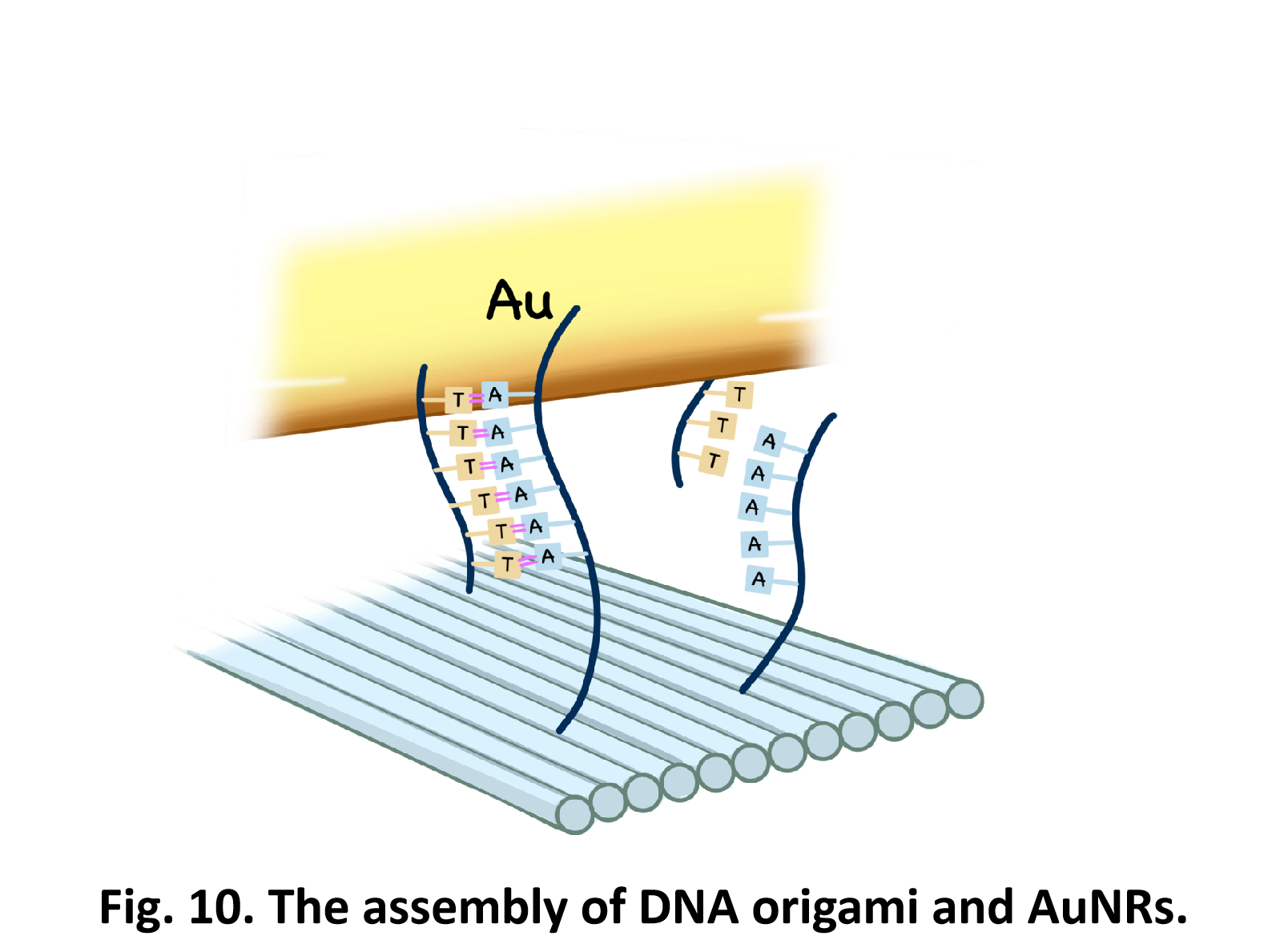
We have functionalized the upper and lower layers of the DNA origami with single strand DNA. These DNA strands can hybridize with the DNA modified on the AuNRs through the Watson-Crick base pairing principle, thereby facilitating the connection of the AuNRs to the DNA origami structure.[13]
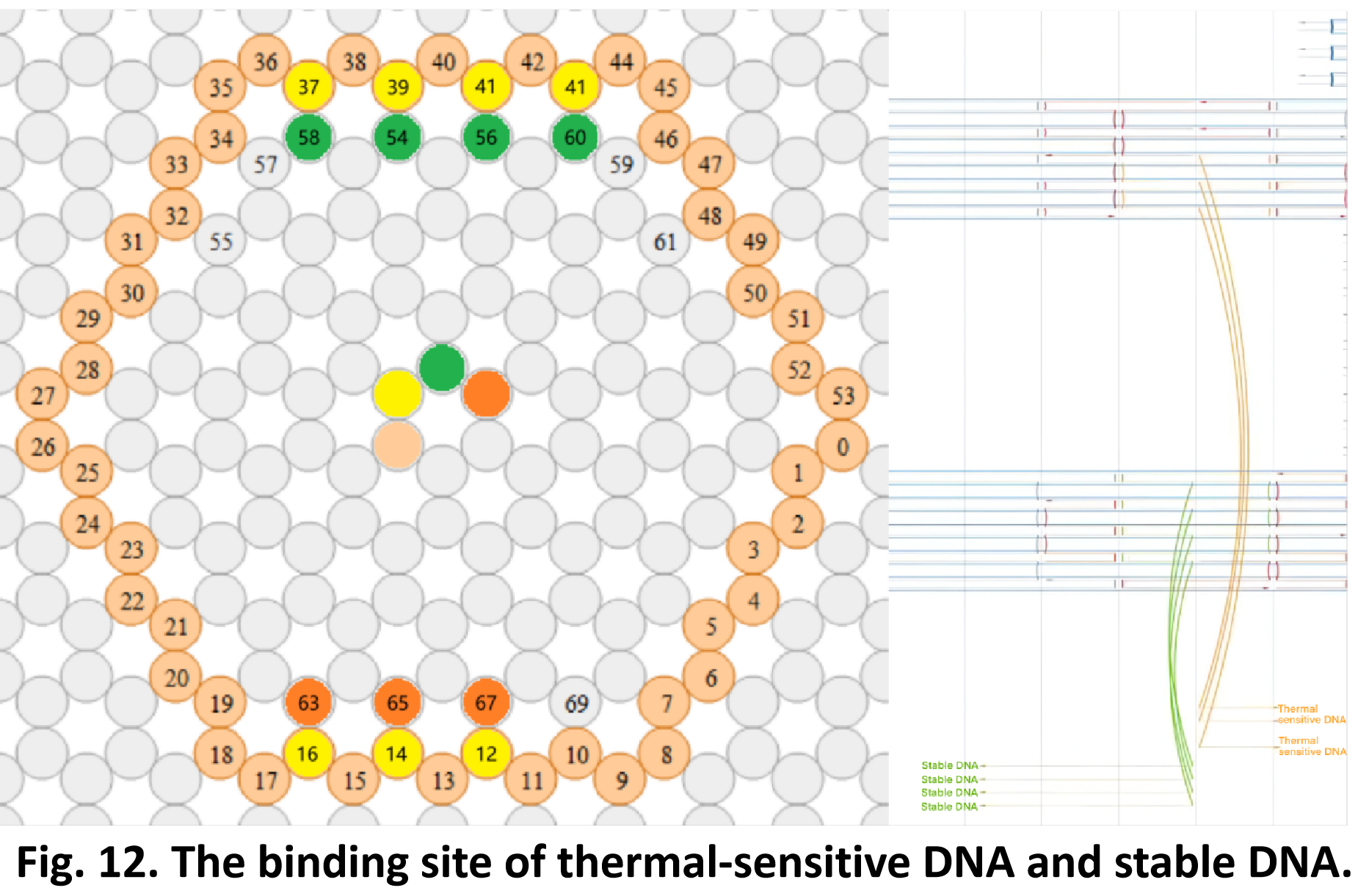
The DNA strand connect to AuNR load on the upper layer of origami is AAAAAAAA. It has only eight bases and a total of three linking sites, and it is a heat-sensitive DNA sequence that can be dehybridized at a AuNR surface temperature of 45℃.[1]
The DNA strand connected to the AuNR loaded on the lower layer of origami is AAAAAAAAAAAAAAAAAAAA. It has a total of 20 bases and four binding sites. It is a relatively stable DNA strand that will not be dehybridized at a AuNR surface temperature of 45℃.[1]


DOARA

See what it can do
So far, We have successfully assembled Doara. Upon exposure to 808 nm near-infrared light, AuNR initiates surface plasmon resonance, which generates heat and induces the dehybridization of DNA on the AuNR surface. This process results in the opening of the Doara. Once the irradiation is ceased, the DNA on the AuNR rehybridizes, leading to the closure of the Doara. Through this mechanism, we have developed a nanostructure that can be remotely controlled by light, showcasing the potential for light-controlled nanodevices in various applications.

References
[1] Johnson JA, Dehankar A, Winter JO, Castro CE. Reciprocal Control of Hierarchical DNA Origami-Nanoparticle Assemblies. Nano Lett. 2019;19(12):8469-8475. doi:10.1021/acs.nanolett.9b02786.
[2] Oberthür D, Achenbach J, Gabdulkhakov A, et al. Crystal structure of a mirror-image L-RNA aptamer (Spiegelmer) in complex with the natural L-protein target CCL2. Nat Commun. 2015;6:6923. Published 2015 Apr 22. doi:10.1038/ncomms7923.
[3] Chen H, Cong X, Wu C, et al. Intratumoral delivery of CCL25 enhances immunotherapy against triple-negative breast cancer by recruiting CCR9+ T cells. Sci Adv. 2020;6(5):eaax4690. Published 2020 Jan 29. doi:10.1126/sciadv.aax4690.
[4] Douglas SM, Marblestone AH, Teerapittayanon S, Vazquez A, Church GM, Shih WM. Rapid prototyping of 3D DNA-origami shapes with caDNAno. Nucleic Acids Res. 2009;37(15):5001-5006. doi:10.1093/nar/gkp436.
[5] Rothemund PW. Folding DNA to create nanoscale shapes and patterns. Nature. 2006;440(7082):297-302. doi:10.1038/nature04586.
[6] Douglas SM, Bachelet I, Church GM. A logic-gated nanorobot for targeted transport of molecular payloads. Science. 2012;335(6070):831-834. doi:10.1126/science.1214081.
[7] Kim DN, Kilchherr F, Dietz H, Bathe M. Quantitative prediction of 3D solution shape and flexibility of nucleic acid nanostructures. Nucleic Acids Res. 2012;40(7):2862-2868. doi:10.1093/nar/gkr1173.
[8] Jabbari A, Sameiyan E, Yaghoobi E, et al. Aptamer-based targeted delivery systems for cancer treatment using DNA origami and DNA nanostructures. Int J Pharm. 2023;646:123448. doi:10.1016/j.ijpharm.2023.123448.
[9] Liu K, Lukach A, Sugikawa K, et al. Copolymerization of metal nanoparticles: a route to colloidal plasmonic copolymers. Angew Chem Int Ed Engl. 2014;53(10):2648-2653. doi:10.1002/anie.201309718.
[10] Zhang X, Servos MR, Liu J. Instantaneous and quantitative functionalization of gold nanoparticles with thiolated DNA using a pH-assisted and surfactant-free route. J Am Chem Soc. 2012;134(17):7266-7269. doi:10.1021/ja3014055.
[11] Chan MH, Chang YC. Recent advances in near-infrared I/II persistent luminescent nanoparticles for biosensing and bioimaging in cancer analysis. Anal Bioanal Chem. 2024;416(17):3887-3905. doi:10.1007/s00216-024-05267-z.
[12] Zhan P, Urban MJ, Both S, et al. DNA-assembled nanoarchitectures with multiple components in regulated and coordinated motion. Sci Adv. 2019;5(11):eaax6023. Published 2019 Nov 29. doi:10.1126/sciadv.aax6023.